
Dendritome Mapping of Genetically-Defined and Sparsely-Labeled Cortical and Striatal Projection Neurons
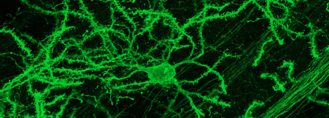
Neuronal morphology is one of the key features for unbiased classification of neuronal cell types in the mammalian brain. Here we propose a novel approach to perform comprehensive brain-wide profiling of the dendritic morphology of genetically-defined neurons in the mouse brain. We have developed an innovative mouse genetic tool, called Mosaicism with Repeat Frameshift (MORF), which enables sparse and stochastic labeling of genetically-defined neurons in mice. MORF reporter mice can label in exquisite detail single neurons from dendrite and spines to axons and axonal terminals at a labeling frequency of 1-5% of a given Cre+ neuronal lineage. We propose to cross our new MORF lines with Cre mouse lines for striatal medium spiny neurons (MSNs) of direct- (D1-MSNs) and indirect-pathways (D2-MSNs), and Layer 5 cortical pyramidal neurons (CPNs) and image the detailed dendritic morphology of thousands of these genetically-defined striatal and cortical neurons (i.e. dendritome). We will digitally reconstruct thousands of MORF-labeled neurons using our novel program called G-Cut and register the brain-wide single neuron morphological data onto a standard reference mouse brain atlas. Reconstructed neurons will subsequently be used for morphology based clustering to define new morphological subtypes. This dendritome data will be disseminated to the Brain Cell Data Center (BCDC) for data integration with those from other BRAIN Initiative Cell Census Network (BICCN) and for data access by the broader neuroscience research community. In addition to the dendritome data generation and analyses, we will further advance our MORF method by generating new MORF reporter mouse lines with logarithmic fold decrease in the Cre-dependent labeling frequencies, which will permit imaging of the complete, brain-wide morphology (i.e. both dendritic and axonal arborizations) of genetically-defined single neurons. Finally, we will develop integrated computer software and hardware (e.g. domain-specific computing) solutions for image processing and neuronal reconstruction, a major bottleneck in analyzing large-scale neuronal morphological datasets. In summary, we will provide rich dendritome information to enable unbiased, morphology-based neuronal cell type classification, and novel mouse genetic tools and computer software and hardware to advance the field of large-scale neuronal morphological studies in the mammalian brain.
Project Leadership
X. William Yang, M.D., Ph.D. (MPI & Contact PI)
Professor, Center for Neurobehavioral Genetics at the Semel Institute for Neuroscience and Human Behavior
Department of Psychiatry & Biobehavioral Sciences
Brain Research Institute
David Geffen School of Medicine at the University of California, Los Angeles
http://yanglab.npih.ucla.edu/?page_id=18
Hong-Wei Dong, Ph.D. (MPI)
Professor of Neurology, Director of Center for Integrative Connectomics,
USC Mark and Mary Stevens Neuroimaging and Informatics Institute
Keck School of Medicine of University of Southern California
http://donglab.loni.usc.edu/
Jason Cong, Ph.D. (Co-PI)
Distinguished Chancellor’s Professor, UCLA Computer Science Department
Director, Center for Customizable Domain-Specific Computing
Director, VLSI Architecture, Synthesis, and Technology (VAST) Laboratory
https://vast.cs.ucla.edu/people/faculty/jason-cong/
Project Data Types
- MORF line will be crossed with D1-Cre (D1-MSNs), D2-Cre (D2-MSNs), and Rpb4-Cre (Layer 5 CPNs) to specifically sparse-label genetically defined neuronal populations.
- For each MORF/Cre line, 10 brains will be collected at P56.
- High-resolution images of D1- or D2-MSNs (about 30,000 to 36,000 neurons per brain) and L5 CPNs (about 10,000 to 20,000 neurons per brain) will be generated using the Andor Dragonfly high-speed confocal system.
- G-Cut will be used to digitally reconstruct the dendritic morphology and individually segment the imaged D1-MSNs, D2-MSNs, and L5 CPNs (5,000 reconstructions per cell type).
- Reconstructed neurons will be registered to the AIBS Common Coordinate Framework (CCF), providing a 3D context of MORF labeled CPNs and MSNs mapped onto a standard atlas.
- Reconstructed and registered neurons will be presented through an online visualization tool similar in its build and functionality to iConnectome viewer.
- The anatomic and dendritic morphological data of the reconstructed neurons will be analyzed and compared to generate unbiased clusters of subtypes within each specific genetically-defined neuronal population.
- New MORF mouse lines with logarithmic decrease in labeling frequency (e.g. 1 in 103 to 105) will be generated to allow visualization of the complete dendritic and axonal morphology of labeled single neurons.
- Integrated computer hardware and software for domain-specific customization for automated image processing and neuronal reconstruction will be developed with Jason Cong’s group.
Related Resources
- Yang Lab: http://yanglab.npih.ucla.edu/